Con un normale microscopio ottico a 400x una cellula sana appare come la foto che segue:
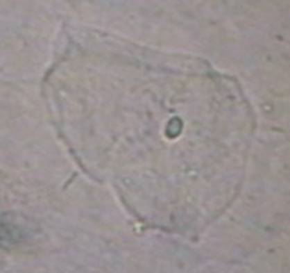
Si notano fondamentalmente tre cose: il nucleo, il citoplasma e la membrana cellulare. Il tutto ha un aspetto “pulito”.
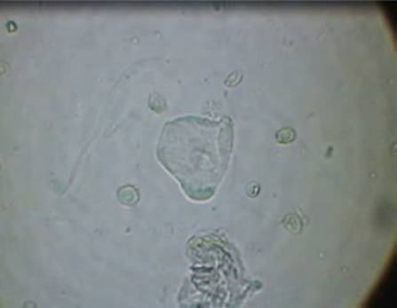
Può accadere invece che, osservando in vivo una qualsiasi cellula del corpo, questa appaia piena di batteri o vescicole come mostrato dalla foto che segue.
Quasi del tutto ignorate fino ad ora, vedremo invece come esse hanno una funzione fondamentale nella formazione della cellula cancerosa.
Queste vescicole, una volta formate, manifestano una naturale tendenza ad aggregarsi, ciò accade sia internamente che esternamente alle cellule.
Quando sono all’interno delle cellule, queste vescicole si aggregano in strutture che occupano spazi abbastanza delineati il più delle volte in periferia e addossate lungo la parete cellulare.
Al di fuori delle cellule invece, queste vescicole si ammassano in aggregati anche molto grandi.
Sui bordi di questi ammassi esterni si formano con facilità delle protuberanze semi sferiche.
Le foto che seguono sono campioni esaminati e fotografati in vivo:
-Fig.1: Una cellula iniziale “pulita”.
-Fig.2: Una cellula che comincia a formare vescicole.
-Fig.3a: Le vescicole che rimangono dentro alla cellula si fondono tra loro e formano un ammassano ben delineato a ridosso della parete cellulare formando in questo esempio delle mezze lune.
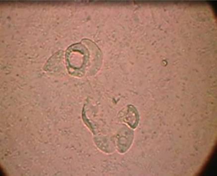
-Fig.3b: Un ammasso di vescicole esterno che presenta in periferia, formazioni circolari.
 Figura 1: Cellula sana


Figura 2: Cellula piena di vescicole


Fig. 3a: Ammasso interno alla cellula. Fig. 3b: Ammasso esterno
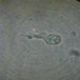
Le piccole formazioni circolari periferiche in fig 3b, abbastanza precocemente mostrano un vorticoso movimento delle vescicole racchiuse al loro interno come mostrato dal filmato che segue.
Il processo é un fenomeno naturale e spontaneo.
In alcuni casi, l’evoluzione ed il passaggio successivo é mostrato dal filmato che segue:
foto di aggregazioni all’interno di una cellula
Da questo momento in poi le cellule che ospitano queste formazioni cominciano pian piano a perdere la loro originale identità.Le nuove formazioni progrediscono a spese della vecchia cellula che un pò alla volta si disgrega e “scompare” come mostrato dalle foto che seguono.

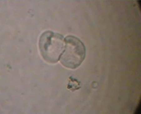
Dalla ulteriore maturazione di tutte queste nuove strutture si forma una cellula particolare: la cellula a forma di clava.
Le foto che seguono ne mostrano alcuni esempi:
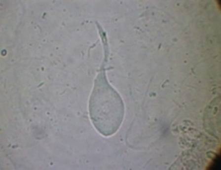

Questa é una cellula molto importante per l’evoluzione del processo canceroso.
Essa non appartiene a nessun tessuto umano.
E’ una cellula completamente nuova.
Precedono la formazione della massa tumorale e quando le ritroviamo nei test di diagnosi precoce sono motivo di preoccupazione appunto per questo motivo.
La loro presenza non ci dice dove si sta formando la massa tumorale ma è il segnale che ci avverte con anticipo che da qualche parte essa si sta formando.
Tutte le modificazioni cellulari fin qui descritte, avvengono nell’organismo in tempi abbastanza lunghi.
Lascio immaginare l’importanza di questa impostazione di ricerca che analizzeremo meglio nelle prossime pagine quando parleremo della diagnosi precoce.
Come si può intuire, abbiamo a disposizione una diagnosi precocissima rispetto ai test tradizionali che hanno invece bisogno della massa tumorale o della cellula cancerosa pienamente sviluppata.
Uno degli obiettivi dell’oncologia classica: “la diagnosi precoce di cancro” diventa in tal modo molto più efficace.
La successiva evoluzione di queste cellule claviformi, fondamentalmente ancora immobili, si manifesta nella loro capacità di diventare mobili con la formazione di chiari ed estesi pseudopodi.
Dal punto di vista diagnostico, quando le cellule cancerose diventano mobili, il processo canceroso é ben avanzato ed esiste già da tempo la massa tumorale.
Le cellule mobili che si formano e che troviamo in un campione da esaminare non hanno però tutte la stessa “gravità”.
Esse possono essere distinte in cellule a lento movimento dalle altre che si muovono invece più speditamente.
{youtube}P0QLibIRMGg{/youtube}
Le cellule a lento movimento non sono cellule cancerose “pericolose”, esse sono giovani e non procurano ancora “danni”.
In questa fase, l’unico problema per il paziente deriva solo dal volume che può raggiungere la massa tumorale.
Vedremo tra breve che differenza c’é tra una cellula cancerosa giovane ed una vecchia e quando e perché essa diventa veramente pericolosa.
Rivediamo tutte le figure che troviamo nei vari stadi fino alla cellula cancerosa mobile:
Cellula sana
La cellula che comincia ad avere la reazione vescicolare.

 c1 c2
c1 c2 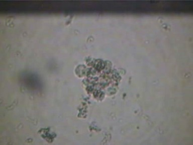 c3
c3
c1-c2) Le vescicole che si riorganizzano all’interno delle cellule
c3) Vescicole che si organizzano all’esterno cellula originaria.

Da entranbe le fasi precedenti si giunge alla prima cellula a forma di clava.

La cellula cancerosa diventa mobile.
Tutte le fasi fino ad ora descritte sono un processo continuo.
Mentre i primi ammassi vescicolari interni ed esterni maturano ed evolvono nel modo che abbiamo appena descritto, altri nuovi se ne formano in tutto il corpo per la reazione vescicolare che avviene in altre cellule “sane” colpite successivamente.
Il tutto avviene con un ritmo continuo, lento ma incessante.
segue con IL PROCESSO CANCEROSO (come evolve la malattia)
–
per contatti telefonici:
Dr. Armando Vecchietti
335 5684474