E’ chiaro che, sapendo riconoscere i vari passaggi fino ad ora descritti e riconoscendo al microscopio le varie modificazioni cellulari, secondo le ricerche del dr. Wilhelm Reich si riesce ad individuare l’insorgere di un processo canceroso anche quando per l’oncologia ufficiale ancora non esiste la benché minima presenza di cellula cancerosa.
Viene da se che potremmo avere a disposizione un’arma incredibilmente potente: la diagnosi precoce.
In questo contesto il sangue gioca un ruolo fondamentale.
Per il fatto che circola in tutto il corpo esso é in grado di riportarci informazioni di prima mano su tutto lo stato energetico dell’organismo.
Qui di seguito vediamo il disegno di due stati energetici che possiamo trovare nel sangue:
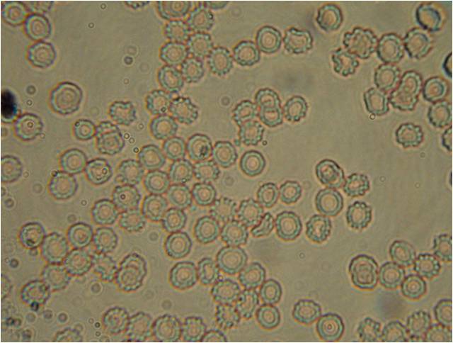
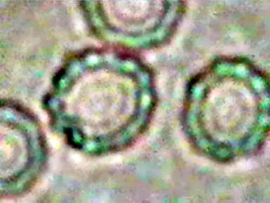
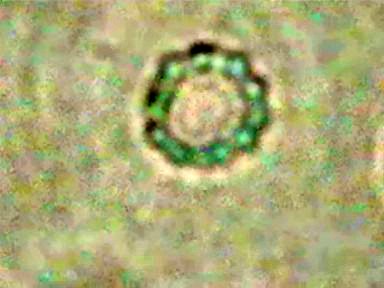
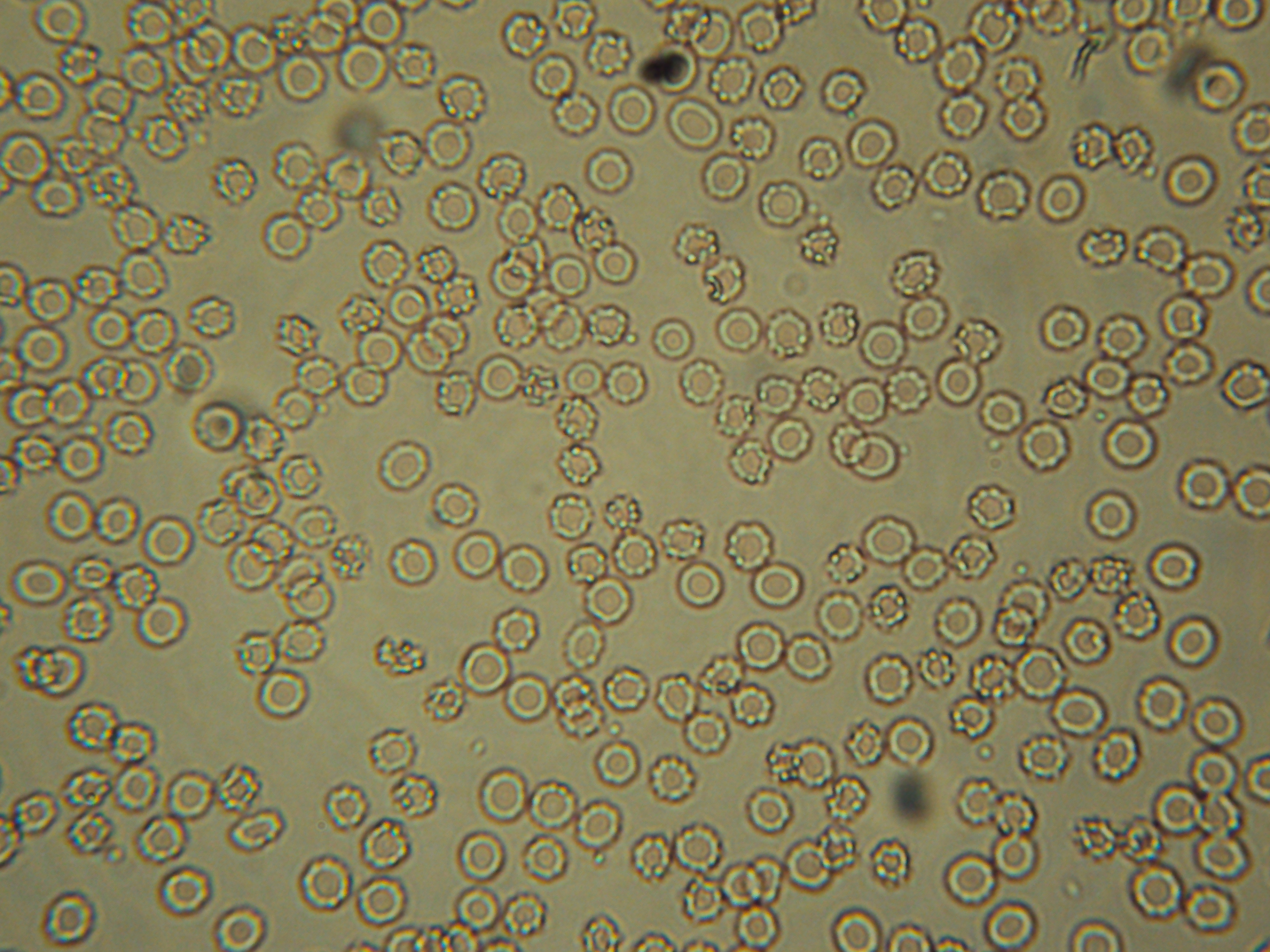
Il sangue sano e carico mostra globuli rossi simili ad una ciambella con delle vescicole incastonate nello stroma come se fossero delle perle.
La reazione vescicolare delle cellule ai bacilli T é in se stessa una reazione sana e corrisponde ad una reazione difensiva che spiegheremo in un’altra occasione.
Anche nei globuli rossi essa corrisponde ad una reazione sana.
Qui le vescicole essendo molto poche non hanno modo di aggregarsi e dare origine alle cellule cancerose come nei tessuti.
Quando si nota la reazione vescicolare all’interno dei globuli rossi questo é il segno che essi sono carichi e capaci di difendere l’organismo.
In un altro contesto spiegheremo come la carica energetica dei globuli rossi può spiegare in modo convincente il fenomeno dell’angiogenesi e come essa non sia uno strumento con il quale il tumore assume nutrimento ma sia una forte reazione di difesa dell’organismo. Comprenderemo facilmente anche quando come e perchè l’angiogenesi si trasforma per l’organismo in un autogol.

Il filmato che segue mostra la reazione del sangue carico energeticamente:
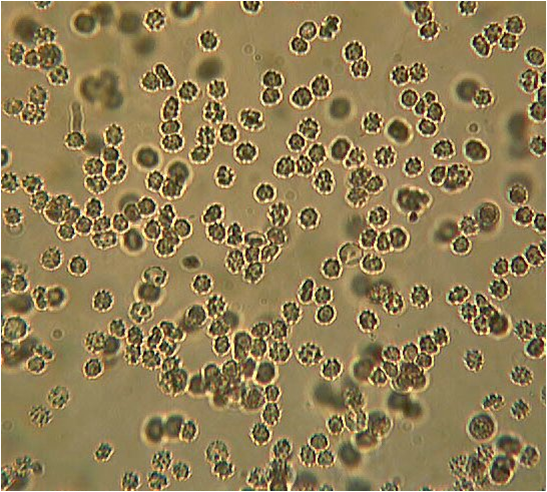
Quando invece i globuli rossi hanno l’aspetto di una sfera spinosa, il sangue é debole e l’organismo é in sofferenza.
Stadi di sviluppo del processo canceroso
Solo a scopo di studio il processo canceroso può essere suddiviso in cinque stadi di sviluppo denominati: Ca1 – Ca2 – Ca3 – Ca4 – Ca5.
Stadio Ca1
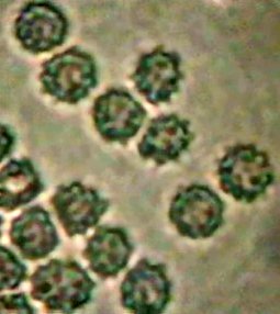
Abbiamo visto come questo stadio é caratterizzato dal danneggiamento dei tessuti mediante il gonfiamento e la reazione vescicolare delle cellule “sane”.
Abbiamo una situazione Ca1 quando le cellule dei tessuti cominciano a mostrare la reazione vescicolare e il sangue presenta la reazione spinosa dei globuli rossi.
Questa prima fase scaturisce da una situazione bioenergeticamente debole delle cellule che compongono i tessuti.
Con un semplice esempio é come se un’azienda in difficoltà (la cellula) cominciasse a “licenziare” i propri dipendenti per difendersi dalla crisi che la attanaglia.
In questa fase, qualsiasi tipo di cellula ispezionabile del corpo osservata in vivo al microscopio, invece di mostrare un citoplasma chiaro si può presentare granulata da vescicole o piena di bacilli T molto piccoli (0,2-0,3 micron).
Ogni volta che i bacilli T sono ritrovati all’interno di una cellula, normalmente essi sono presenti anche nei fluidi e quindi circolanti nell’organismo.
La cellula colpita generalmente tende anche ad arrotondarsi a causa della contrazione.
Nelle cellule epiteliali questa contrazione è chiaramente osservabile negli angoli che diventano arrotondati e nella graduale perdita della forma pentagonale.
In questo stadio di gonfiamento, la tendenza alla reazione vescicolare rapida e la formazione di bacilli T è uno dei segnali più importanti dell’imminente sviluppo canceroso.
Quando a questo quadro si associa anche il test del sangue che mostra una aspetto spinoso dei globuli rossi, possiamo fare una diagnosi di Ca1.
Per l’oncologia classica, in questo stadio non esiste ancora nessun tipo di cancro in quanto non è ritrovata nessuna cellula tumorale.
Nei casi in cui il danneggiamento è chimico o traumatico è ragionevole pensare che lo stimolo canceroso avvenga sempre e comunque attraverso la formazione di bacilli T che scaturiscono dalla morte dalle cellule dei tessuti bio-energeticamente più deboli.
I test che abbiamo a disposizione per il sangue sono di due tipi.
La grossa differenza tra questi ed i comuni test della medicina tradizionale consiste solo nel fatto che i primi sono bioenergetici i secondi sono test biochimici.
La produzione di grosse vescicole blu all’interno dei globuli rossi sono il segno di una loro forte carica.
Quando invece i globuli rossi mostrano un aspetto spinoso la loro carica è molto scarsa e così quella dell’intero organismo.
Essi ci avvertono quindi anche con decenni di anticipo se l’organismo sta scivolando o meno verso una patologia cancerosa.
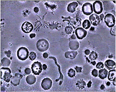
Le seguenti foto sono cellule Ca1.
Stadio Ca2
Questo stadio é caratterizzato da uno stato infiammatorio acuto.
La reazione vescicolare delle cellule continua.
I bacilli T (gram negativi) continuano ad essere presenti e non subiscono successive modificazioni.
Essi sono ritrovati e mantengono le stesse caratteristiche in ogni successivo stadio e per tutta la durata del processo canceroso.
Abbiamo visto che la loro presenza è mortale per l’organismo.
Sono loro che spingono sempre più velocemente le cellule sane dei tessuti verso la ormai conosciuta reazione vescicolare.
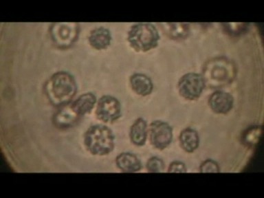
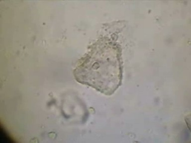
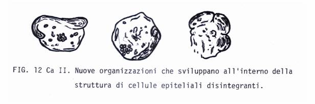
In questo stadio si vede la prima forma evolutiva delle vescicole che da omogeneamente distribuite all’interno delle cellule, cominciano ad aggregarsi in prossimità della parete cellulare.
Queste nuove aggregazioni perdono il loro grossolano aspetto vescicolare, diventano ammassi sempre più delineati ed evolvono a spese della vecchia cellula originaria.
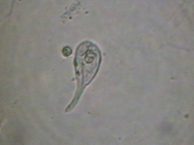
In alcuni casi, sin da subito si possono notare le prime forme affusolate, ovoidali e caudate.
A volte la porzione restante della cellula colpita si frantuma in piccoli detriti o in vescicole ancora più piccole. Le immagini che seguono sono cellule Ca2.
Di preferenza le proliferazioni infiammatorie raggiungono le ghiandole.
Ad un esame microscopico di questi tessuti, sia nel preparato vivo che in quello da autopsia, la medicina tradizionale non riconosce ancora nessuna traccia di cellule carcinomatose.
Quando vediamo che all’interno di una cellula le vescicole cominciano a riorganizzarsi, possiamo fare una diagnosi di Ca2.
Stadio Ca3
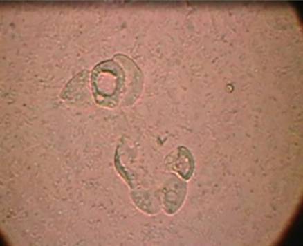
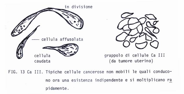
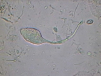
Questo stadio é caratterizzato dalla presenza di cellule isolate a forma di fuso, di clava o caudate spesso presenti in proliferazioni infiammatorie diventate croniche.
E’ in questo stadio che la medicina ufficiale riconosce a volte le prime cellule cancerose vere e proprie.
All’inizio queste nuove cellule possono mostrare una struttura altamente vescicolare, striata e sono vivacemente luminose.
Esse hanno forma affusolata, caudata, ovale o rotonda.
Mostrano grandi variazioni in grandezza.
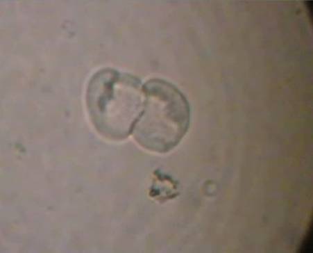
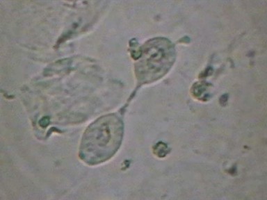
Si moltiplicano rapidamente e possono essere osservate in fase di divisione.
Va ricordato che le osservazioni devono sempre essere eseguite in vivo.
A livello vaginale tutte queste cellule sono normalmente eliminate con la secrezione ma nell’utero come negli altri organi pelvici la reazione vescicolare sulle cellule restanti é un processo continuo.
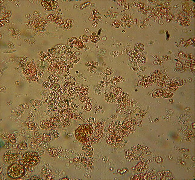
Quando le troviamo in altri organi, queste cellule formano densi grappoli, formano la massa tumorale e sono responsabili delle infiltrazioni nel circostante tessuto.
Questo stadio è denominabile carcinomatoso ed è caratterizzato da processi non rintracciabili in tessuti infiammati o sani.
Le nuove formazioni cellulari si distinguono per la loro colorabilità biologica naturale straordinariamente intensa presente nei diversi gradi di organizzazione.
Al contrario, la cromo sezione morta di un campione non ci rivela ancora nulla.
In questo stadio anche una sola cellula sana si può trasformare in una struttura precancerosa a forma di fuso ma di solito si vedono disgregarsi in vescicole anche cellule sane contigue.
Queste confluendo in un unico mucchio vescicolare generano nuove cellule cancerose da un ammasso che hanno formato in comune.
In genere sono cellule a pessima motilità non eccessivamente “dannose”.


Cellula a lento movimento
La diagnosi di Ca3 può essere fatta quando nel preparato in vivo, sia pure in modo anche isolato, compaiono sia la decomposizione vescicolare che le nuove forme a fuso.
Va sottolineato che i bacilli T sono responsabili unicamente dell’innesco dello stadio canceroso.
Le cellule cancerose, una volta scaturite, si sviluppano ed evolvono in modo completamente autonomo cioè indipendente dai fattori che le hanno generate.
La “penetrazione distruttiva” delle cellule cancerose va in gran parte attribuita non alla cellula cancerosa in se stessa ma piuttosto alla medesima reazione vescicolare che si attua anche nelle cellule adiacenti.
In questo modo la cellula cancerosa perde di importanza per quanto riguarda il danneggiamento tessutale che precede la sua formazione in altri tessuti.
La cellula cancerosa non é la causa del cancro, non si forma come portatrice di malattia e di morte, essa è solo la conseguenza della riorganizzazione vescicolare delle cellule.
Essa diventa un pericolo mortale soltanto alla fine del suo ciclo vitale quando morendo, va in putrefazione come qualsiasi altro organismo.
Va precisato che la riorganizzazione delle vescicole in cellule cancerose e il loro proliferarsi in tumore maligno è un processo autonomo e indipendente dall’azione dei fattori che innescano il processo.
Le Ca3 sono cellule claviformi, possono essere anche a grappolo e sono quelle che vanno a generare la prima massa tumorale.
Il disegno e le foto che seguono rappresentano lo stadio Ca3.
In questo stadio non é difficile imbattersi in cellule che si possono vedere in fase di divisione.
Stadio Ca4
Lo stadio di Ca4 è caratterizzato da cellule cancerose completamente mature.
Le cellule Ca3 continuando ad evolvere mostrando un aspetto unificato, ialino, di forte contrasto.
Alcune di queste cellule caudate possono sviluppare anche flagelli e motilità.
Lo stadio Ca4 é caratterizzato dalla presenza di cellule mobili con movimenti ameboidi vivaci e sono il prodotto maturo e finale delle precedenti cellule a forma di fuso.
Il filmato che segue sono cellule Ca4:
Filmato Cellule Ca4
In altri casi le cellule che hanno assunto una forma rotonda si allungano e diventano mobili attraverso la formazione di estesi e chiari pseudopodi.
A volte gli pseudopodi possono essere di tipo filamentoso.
Altre volte attorno ad ogni ammasso di vescicole si può sviluppare una membrana che lo avvolge e lo racchiude, in questo caso possono scaturire anche dei protozoi.
Questo cellule rappresentano lo stadio Ca4.
La cancerologia classica ritiene che esse siano invece dei “parassiti”.
Se l’organismo non morisse prima, queste formazioni si trasformerebbero in amebe vere e proprie.
In questo stadio la malignità del tumore dipende dal grado di maturità delle cellule cancerose e dalla rapidità con cui esse e le parti del tessuto distrutto vanno in decomposizione.
Stadio Ca5
Questo stadio è caratterizzato dalla morte e dalla decomposizione putrida delle cellule che compongono i tessuti.
E’ la fase terminale della malattia.
Quando al microscopio si ritrovano cellule caudate e capaci di movimento, il cancro è ben avanzato.
Lo stadio Ca5 è caratterizzato da necrosi.
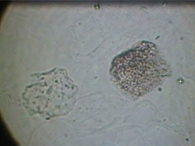
L’immagine microscopica della fase Ca5 mostra la presenza di molti detriti, di frammenti di cellule, di vescicole, batteri e ad ingrandimenti più forti, dei soliti corpuscoli T.
Si notano bene i pezzi delle cellule del Ca2 ancora presenti, essi mostrano una granulazione scura.
Lo stadio Ca5 è determinato e caratterizzato quasi esclusivamente dalle cellule cancerose che giunte alla fine del loro ciclo vitale, muoiono.
Esso è paragonabile alla necrosi post morte dei tessuti.
Le cellule cancerose hanno vita breve ed appena morte vanno in decomposizione.
Ne risulta una batteriemia ed una tossicemia generalizzata dell’organismo.
Quando il tumore non pregiudica funzioni vitali la morte avviene per putrefazione tossica generalizzata.
Questo spiega perché, giunta alla fine, la malattia generalmente peggiora rapidamente e sopraggiunge la morte.
A questo stadio qualsiasi terapia diventa inutile.
Riepilogo del processo canceroso
prima fase
1) Reazione vescicolare delle cellule come reazione di difesa.
2) Formazione delle cellule cancerose dagli ammassi vescicolari e loro proliferazione.
3) Formazione della massa tumorale.
seconda fase
4) Morte e decomposizione putrida delle cellule che compongono la massa tumorale.
5) Massiccia, progressiva ed accelerata putrefazione del sangue e dei tessuti, produzione di detriti e batteri della putrefazione e bacilli T.
6) Cachessia, putrefazione generalizzata di tutto l’organismo e conseguente morte.
In un individuo con un alto livello energetico la produzione di vescicole pur verificandosi ugualmente avviene in maniera più lenta.
Il tessuto vecchio che si disgrega viene riassorbito ed alla stessa velocità se ne forma di nuovo.
I detriti sono eliminati tanto rapidamente quanto essi sono formati.
In un paziente con un basso livello energetico si osserva un quadro completamente diverso:
Nel Ca1 e Ca2 all’inizio troviamo un processo di contrazione ed una reazione vescicolare più rapida.
La ricarica dell’individuo che si trova in queste condizioni é ancora un processo possibile e reversibile.
Anche nella fase di Ca3 (tumori di prima necrosi) il processo é reversibile.
Nei casi Ca4 e Ca5 il processo diventa irreversibile dal punto di vista del recupero ma l’espansione provocata dalla ricarica energetica riduce il dolore e può prolungare la vita migliorando la qualità della vita ed il paziente è più disponibile a lottare contro la malattia.
Quando un paziente comincia a caricarsi energeticamente, nel suo organismo si verificano una serie di cambiamenti.
L’individuo acquista un aspetto più vivo, meno affaticato e percepisce una soggettiva sensazione di benessere.
Organismo in toto:
Reazione sana: Elasticamente eretto, tono buono, assenza di spasmi e cloni. Sensazione di vigore. Capacità di provare piacere.
Reazione malata: Contratto, flaccido o ipotonico. Spasmi e cloni. Sensazione di debolezza. Incapacità di provare piacere. Ansietà del piacere.
Pelle:
Reazione sana: Calda e irrorata dal sangue. Turgore buono, rosa o abbronzata, capace di produrre sudore caldo.
Reazione malata: Fredda e viscida. Poco irrorata. Turgore povero, pallida e livida. Sudore freddo.
Muscolatura:
Reazione sana: Rilassata, capace di alternare tensione e rilassamento, forte. Nessun muscolo corrazzato, vivace peristalsi, nessuna costipazione o emorroidi.
Reazione malata: Tensione cronica o flaccida e atrofica. Spesso grasso eccessivo. Corazzatura muscolare generalizzata. Costipazione ed emorroidi.
Espressione del volto:
Reazione sana: Vivacemente variabile.
Reazione malata: Rigida come una maschera, espressione morente.
Sangue:
Reazione sana: Reazione B al test dell’autoclave. Eritrociti tesi, pulsanti, che mostrano un forte ed ampio margine energetico. Bassa disintegrazione in fisiologica. Nessun bacillo T in coltura.
Reazione malata: Reazione T al test dell’autoclave. Eritrociti contratti e piccoli, non pulsanti, che mostrano corpuscoli T. Energeticamente debole, rapida disintegrazione in fisiologica. La coltura mostra streptococchi, bacilli T o stafilococchi.
Sistema cardio-vascolare:
Reazione sana: Polso regolare, calmo e forte. Pressione del sangue normale.
Reazione malata: Polso irregolare vivace o debole. Pressione anormalmente alta o bassa.
Tessuti (cellule epiteliali, tessuti ottenuti da biopsia):
Reazione sana: Turgore vigoroso. Nessuna formazione di vescicole (bioni) in soluzione KCl.
Reazione malata: Turgore debole, contratto. Strutture bioniche o rapida disintegrazione vescicolare (bionica) in KCl.
Occhi:
Reazione sana: Splendenti, vivaci, reazione pupillare vivace. Non sporgenti nè incavati.
Reazione malata: Depressi, lontani. Reazione pupillare lenta, spesso midriaca. Occhi sporgenti o incavati.
Respirazione:
Reazione sana: Piena espirazione con una pausa al termine di essa, libera pulsazione del torace. Sensazione di piacere genitale dopo ogni respirazione.
Reazione malata: Inibita ed incompleta espirazione con una pausa dopo l’inspirazione. Fissazione in una cronica attitudine di inspirazione (ansietà). Nessuna sensazione di piacere dopo l’espirazione.
Orgasmo:
Reazione sana: Si verifica regolarmente. Piena convulsione del corpo. Nessuna stasi sessuale.
Reazione malata: Assente o disturbato. Cronica stasi sessuale.
segue ANGIOGENESI
–
per contatti telefonici:
Dr. Armando Vecchietti
335 5684474